Department Lead

Dr. Jaideep Sharma
M.B.B.S., M.S., DNB, FAICO, FICO (U.K.), FAEH | Vitreoretina Expert
Recognized as a leading retina specialist in Jaipur, Dr. Jaideep Sharma brings rigorous surgical protocols from the prestigious Aravind Eye Hospital. He specializes in the management of complex posterior segment pathologies, executing emergency pars plana vitrectomies, complex macular hole repairs, and advanced diabetic retinopathy laser interventions with pinpoint precision.
Why Choose Prasan Nethralaya for Retinal Care?
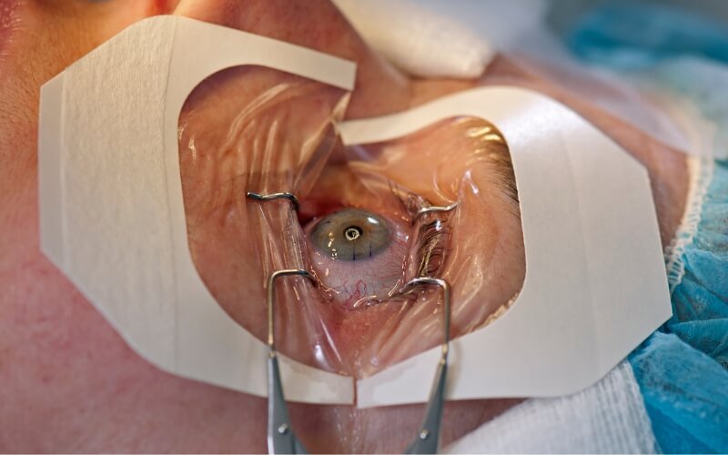
Retinal surgery is the most delicate discipline in ophthalmology. We combine master-class surgical training with cutting-edge micro-instrumentation.
Emergency Surgical Readiness
Conditions like Retinal Detachment require urgent action. Our operation theaters are equipped and our team is primed to perform emergency sight-saving procedures.
MIVS (Micro-Incision Vitrectomy)
We utilize 25-gauge and 27-gauge sutureless surgical techniques. Smaller incisions mean dramatically faster recovery, less post-operative pain, and better visual outcomes.
Comprehensive Diabetic Care
We offer a holistic approach to Diabetic Retinopathy, seamlessly integrating laser therapies, Anti-VEGF injection protocols, and long-term disease management.
Your Retina Care Journey
Navigating retinal disease can feel overwhelming. We break down your care into a clear, predictable pathway to preserve your sight.
Deep Diagnostics
Pupil dilation followed by high-resolution OCT scans to map the microscopic layers of your macula and retina.
Clinical Consultation
Dr. Jaideep reviews your scans with you in real-time, explaining the pathology and laying out treatment options.
Targeted Intervention
Execution of medical therapy (Injections/Laser) or advanced surgical intervention (Vitrectomy) to halt disease.
Chronic Management
Ongoing, scheduled monitoring to prevent recurrence, particularly for chronic conditions like Diabetes and ARMD.
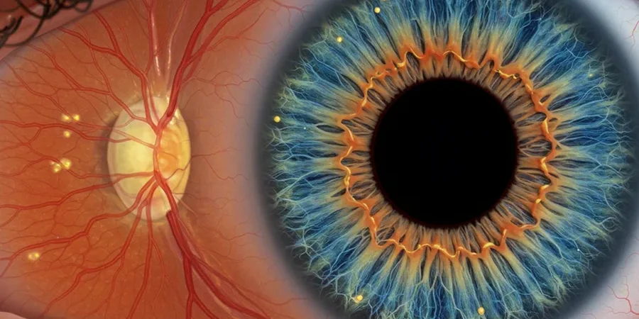
Understanding Retinal Health
Think of the eye like a camera. The cornea and lens focus the light, but the retina is the high-resolution film at the back of the eye that captures the picture. The central part of the retina, called the macula, is responsible for sharp, detailed, central vision required for reading and driving.
Because the retina is an extension of the brain and relies on a microscopic network of blood vessels, it is highly susceptible to systemic diseases like diabetes, hypertension, and the natural aging process. Damage to this tissue is often painless, making regular dilated eye exams crucial.
Red Flag Symptoms: When to See a Retina Specialist
- • Sudden appearance of dark floaters or "cobwebs"
- • Flashes of light in your peripheral vision
- • A dark curtain or shadow falling across your vision
- • Distortion of straight lines (metamorphopsia)
- • Sudden, unexplained blurring or loss of central vision
Core Retinal Pathologies
Diabetic Retinopathy
Prolonged high blood sugar weakens retinal blood vessels, causing them to leak fluid or bleed. Leads to severe vision loss if untreated.
Retinal Detachment
A critical medical emergency where the retina pulls away from supportive tissue. Causes permanent blindness without rapid surgery.

Macular Degeneration (ARMD)
The leading cause of severe vision loss in patients over 60, destroying the macula and erasing central vision capabilities.

Macular Hole
A small break in the macula located at the center of the eye's light-sensitive tissue, causing blurry and distorted central vision.

ROP (Infant Retina)
A potentially blinding eye disorder affecting premature infants. Abnormal blood vessels grow and can lead to detachment.

Retinal Vein Occlusion
A blockage of the small veins that carry blood away from the retina, often causing sudden blurring or vision loss in one eye.
Advanced Retinal Technology
We arm our surgeons with the highest caliber diagnostic imaging and micro-surgical systems to visualize and repair the microscopic layers of your eye.
Optical Coherence Tomography (OCT)
A non-invasive imaging test that uses light waves to take highly precise cross-section pictures of your retina, allowing us to see each distinct layer and measure its thickness to detect fluid or swelling.
Fundus Fluorescein Angiography (FFA)
A specialized diagnostic procedure where a fluorescent dye is injected into the bloodstream. A special camera tracks the dye as it travels through the retinal blood vessels, instantly highlighting leaks, blockages, or abnormal growth.
Constellation® Vision System
The global gold standard machine for Vitreoretinal surgery. It provides Dr. Jaideep with absolute control over fluid dynamics, lighting, and cutting speed during complex procedures like pars plana vitrectomy.
Patient
Questions.
Clinical clarity regarding posterior segment procedures, recovery, and long-term care.
Early signs include a sudden increase in eye floaters, flashes of light in your peripheral vision, or a dark curtain-like shadow moving across your visual field. This is a medical emergency requiring immediate attention from a retina specialist to prevent blindness.
While the structural damage caused by diabetic retinopathy cannot always be fully reversed, early intervention with laser photocoagulation or Anti-VEGF injections can effectively halt the progression of the disease and prevent further, irreversible vision loss.
The procedure is virtually painless. At Prasan Nethralaya, we apply powerful topical anesthetic drops to numb the eye completely before administering the intravitreal injection. Patients may feel slight pressure, but no sharp pain.
Initial recovery takes 2 to 4 weeks. If a gas bubble was used during surgery to hold the retina in place, you may need to maintain a strict face-down head position for several days to a week. Full visual stabilization can take several months.
All diabetic patients should undergo a comprehensive dilated fundus examination at least once a year. If signs of diabetic retinopathy are detected, Dr. Jaideep may recommend screenings every 3 to 6 months to monitor disease progression.
Protect Your Vision Today
Retinal diseases often present no early pain or warning signs. Schedule a comprehensive diagnostic evaluation with our retina specialists.
Book Consultation