Understanding ROP
Retinopathy of Prematurity (ROP) is a potentially blinding eye disorder that primarily affects premature infants. In a full-term pregnancy, the blood vessels of the retina (the light-sensitive layer at the back of the eye) finish developing just before birth. When a baby is born too early, these vessels have not yet reached the edges of the retina.
After birth, the normal development of these vessels can be disrupted. Instead of growing normally, the eye may begin to grow abnormal, fragile blood vessels. These vessels can leak and cause scar tissue to form. If the scar tissue shrinks, it can pull the retina away from the back of the eye, leading to a retinal detachment and permanent blindness. Because babies cannot tell us if their vision is blurry, specialized screening is the only way to detect and treat ROP in time.
Mandatory Screening Criteria
Under the "Vision 2020" guidelines, the following infants must undergo ROP screening:
- • Birth weight of 1500 grams or less.
- • Gestational age of 32 weeks or less.
- • Infants with a birth weight 1500g-2000g with an unstable clinical course.
- • Any infant receiving prolonged oxygen therapy in the NICU.
The Diagnostic & Treatment Protocol
At Prasan Nethralaya, we provide comprehensive bedside screening in the NICU and follow-up examinations in our specialized pediatric retina suite. ROP is staged by severity (1 to 5) and by Zone (location in the eye).
1. The Digital Screening (RetCam)
We utilize advanced indirect ophthalmoscopy and, where available, wide-field digital imaging (RetCam) to document the peripheral retina. This allows us to track the growth of vessels week by week. The exam is performed after dilating the baby's pupils with mild pediatric drops.
2. Diode Laser Photocoagulation
If ROP reaches "Threshold" or "Type 1" severity, immediate treatment is required—usually within 48 to 72 hours. The gold standard is Laser Photocoagulation. We use a specialized laser to treat the peripheral retina that has no blood vessels. This stops the production of the growth factors that cause abnormal vessels to grow, effectively "cooling down" the disease and preventing detachment.
3. Intravitreal Anti-VEGF Injections
In certain aggressive "Plus" cases or "Zone I" ROP (where the disease is very close to the center of vision), we may administer a microscopic injection of Anti-VEGF medication. This drug rapidly halts the progression of abnormal vessel growth. This is often used as a bridge to allow the eye to stabilize or as a primary treatment in very sick infants.
4. Vitreoretinal Surgery
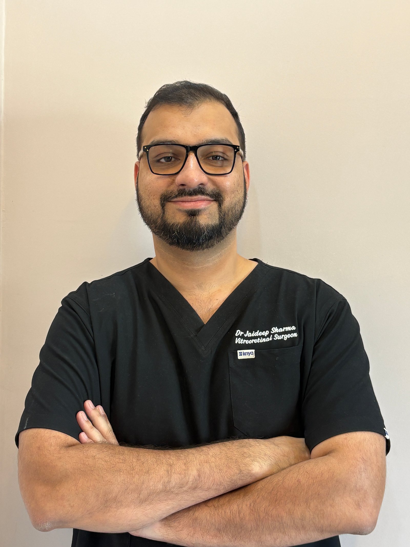
If ROP progresses to Stage 4 or 5 (partial or total retinal detachment), complex Pediatric Vitrectomy is required. Dr. Jaideep Sharma specializes in these microscopic maneuvers to remove scar tissue and reattach the infant retina. However, the goal of our screening program is to catch and treat ROP long before this stage is reached.
The Critical "Rule of 30 Days"
The most important factor in saving a baby's vision is timing. The first ROP exam must be performed no later than 30 days after birth. If the baby was born extremely premature (before 28 weeks), the exam should happen even earlier. ROP is a time-bound emergency; once the retina detaches in Stage 5, the visual prognosis is extremely poor. Following Dr. Jaideep's screening schedule is the most vital responsibility of the parents after NICU discharge.