Department Lead

Dr. Neelam Sharma
Cornea & Anterior Segment Specialist
A master of the anterior segment, Dr. Neelam leads the regional charge in advanced keratoconus management, precision C3R cross-linking, and intricate corneal transplantation procedures. Her clinical protocols are designed to restore flawless optical clarity to compromised eyes.
Why Choose Prasan Nethralaya for Corneal Care?
The cornea is less than a millimeter thick but consists of five distinct layers. We bring absolute microscopic precision to treating complex surface diseases.
Lamellar Grafting Expertise
We prioritize partial-thickness transplants (DALK, DMEK, DSAEK). By surgically replacing only your diseased corneal layers and preserving the healthy tissue, we drastically reduce rejection rates and accelerate healing.
Emergency Ulcer Management
Corneal ulcers can melt through the eye in hours. We provide rapid diagnostic scraping and instantly deploy custom-fortified antimicrobial drops to halt sight-threatening infections.
Proactive Keratoconus Care
We do not wait for your vision to fail. Using sub-clinical topographic screening, we detect keratoconus early and perform C3R cross-linking to structurally freeze the cornea before permanent distortion occurs.
Your Cornea Care Journey
From diagnostic mapping to post-surgical optical rehabilitation, we guide you through every step of restoring your visual surface.
Topographic Profiling
We capture a 3D elevation map and perform an endothelial cell count to evaluate the exact structural integrity of your cornea.
Clinical Diagnosis
Dr. Neelam identifies exactly which of the five corneal layers is diseased, inflamed, or structurally failing.
Targeted Intervention
Executing precise medical therapy, UV cross-linking, or coordinating donor tissue for specialized lamellar transplantation.
Optical Rehabilitation
Post-operative management, including selective stitch removal and specialty contact lens fitting to maximize visual clarity.
Understanding the Anterior Segment
The cornea is the crystal-clear, dome-shaped window at the very front of your eye. It is incredibly powerful, providing about 65% to 75% of your eye's total focusing power. Because it must remain perfectly transparent to allow light to pass through to the retina, it contains no blood vessels. Instead, it relies entirely on tears and the aqueous fluid within the eye for its nourishment.
When the cornea is damaged by trauma, severe infection (ulcers), or progressive diseases like Keratoconus or Fuchs' Dystrophy, it loses its smooth shape and crystal clarity. Light entering the eye becomes scattered and distorted, leading to severe visual impairment. Unlike simple refractive errors, these conditions cannot be fixed with standard glasses and require highly specialized medical and surgical intervention.
Red Flag Symptoms: When to Seek Urgent Care
- • Extreme sensitivity to light (Photophobia)
- • A constant, painful sensation of sand in the eye
- • A visible white or grey patch on the clear part of the eye
- • Rapidly worsening blurry or distorted vision
- • Severe eye redness accompanied by sharp pain
Comprehensive Corneal Interventions

Keratoconus Treatment
A progressive disease where the cornea thins and bulges into a cone shape, causing severe distortion. We utilize advanced topography to detect and halt progression early.
Corneal Transplantation
For severely scarred or diseased corneas, surgical replacement with healthy donor tissue is required to restore functional vision and optical clarity.

Corneal Ulcers
Severe bacterial, fungal, or viral infections that create an open sore on the cornea. This is an absolute medical emergency requiring immediate sterilization.

Severe Dry Eye Syndrome
Chronic lack of sufficient lubrication leading to extreme discomfort, burning, and potential scarring of the corneal surface if left unmanaged.

Pterygium Surgery
A fleshy, wedge-shaped growth of conjunctival tissue that aggressively invades the cornea, causing chronic redness and astigmatism.

Corneal Dystrophies
Genetic disorders, such as Fuchs' Endothelial Dystrophy, where abnormal material accumulates in the cornea, causing progressive fluid buildup and cloudy vision.
Advanced Cornea Technology
To treat the cornea safely, we must measure it flawlessly. Our diagnostic suite provides sub-micron mapping of the ocular surface.
Pentacam HR Topography
The gold standard for diagnosing Keratoconus. It captures a 3D elevation map of the front and back of your cornea, calculating exact tissue thickness at thousands of points in just a few seconds.
Specular Microscope
Essential for diagnosing Fuchs' Dystrophy and evaluating transplant viability. This device takes high-magnification photographs of the endothelium to literally count the number of healthy cells on the back of your cornea.
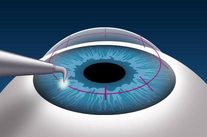
UV-X System for C3R
The advanced medical device used during Corneal Cross-Linking. It delivers a highly calibrated dose of Ultraviolet-A light to activate riboflavin drops, creating new structural bonds to freeze the progression of Keratoconus.
Patient
Questions.
Clinical clarity regarding keratoconus progression, complex transplants, and corneal surface health.
Early signs often include severe light sensitivity (photophobia), a constant feeling that something is in your eye (foreign body sensation), sharp pain, cloudy or hazy vision, and seeing halos around lights at night.
Keratoconus cannot be reliably diagnosed with a standard vision chart early on. It requires advanced Corneal Topography (like a Pentacam scan) to create a 3D elevation map of the cornea to detect microscopic thinning and abnormal bulging.
A full-thickness transplant (PK) replaces all layers of the cornea. A partial-thickness transplant (like DALK or DSAEK) surgically replaces only the specifically diseased layers while leaving your healthy tissue intact, resulting in much faster recovery and a significantly lower risk of tissue rejection.
Yes. Chronic, severe dry eye syndrome deprives the cornea of essential lubrication, oxygen, and nutrients. Over time, this constant friction can lead to painful corneal abrasions, recurrent ulcers, and permanent scarring that severely affects your vision.
A corneal ulcer is a medical emergency usually caused by a severe bacterial, viral, or fungal infection (often linked to improper contact lens use). Treatment requires immediate, aggressive application of fortified antimicrobial eye drops to sterilize the eye and prevent the ulcer from perforating the cornea.
Restore Your Visual Surface.
Whether managing severe dry eye or requiring advanced topographic mapping for Keratoconus, secure your comprehensive corneal evaluation today.
Book Corneal Evaluation